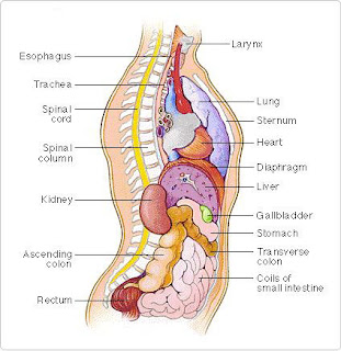
Body/Torso -- Side View
Your torso consists of two parts — the chest and the abdomen. The chest contains your heart and lungs; your abdomen contains the digestive and urinary systems. Your chest and abdomen are separated by a dome-shaped sheet of muscle called the diaphragm.
Hand -- Carpal Tunnel
The carpal tunnel is the area under a ligament (a tough, elastic band of tissue that connects bones and organs in place) in front of the wrist. The median nerve, which passes through the carpal tunnel, supplies the thumb side of the hand. Repetitive movements of the hand and wrist can cause inflammation of structures (such as tendons and their coverings) that surround the median nerve. The inflammation may compress this nerve, producing numbness, tingling, and pain in the first three fingers and the thumb side of the hand - a condition known as carpal tunnel syndrome.
Brain -- Effects of Stroke
Your brain has three main components - the cerebrum (which consists of the left and right cerebral hemispheres), the cerebellum, and the brain stem. The cerebral hemispheres of the brain make up the largest part of your brain. The cerebellum is the structure located behind the brain stem, and the brain stem is the lowest section of the brain and is connected to the spinal cord.
Region of the Cerebrum Damaged by Stroke | Signs and Symptoms |
Wernicke's area (central language area) | Difficulty speaking understandably and comprehending speech; confusion between left and right; difficulty reading, writing, naming objects, and calculating |
Broca's area (speech) | Difficulty speaking and, sometimes, writing |
Parietal lobe on the left side of the brain | Loss of coordination of the right arm and leg |
Facial and limb areas of the motor cortex on the left side of the brain | Paralysis of the right arm and leg and the right side of the face |
Facial and arm areas of the sensory cortex | Absence of sensation in the right arm and the right side of the face Optic radiation Loss of the right half of the visual field of both eyes |
Glossary
Brain stem
Mainly controls unconscious vital functions such as blood pressure and breathing
Broca's area
Controls speech
Cerebellum
Maintains posture, balance and coordination of movement
Gustatory area
Controls the sense of taste
Left cerebral hemisphere
Together with right cerebral hemisphere, controls most conscious and mental activities
Left middle cerebral artery
A major source of blood supply to the brain
Motor cortex
Sends instructions to muscles to cause voluntary movements
Optic radiation
Tract of nerve fibers involved in vision
Parietal lobe
Involved in sensations of pain and touch, spatial orientation, and speech
Prefrontal cortex
Provides ability to plan, reason, concentrate, and adjust behavior
Premotor cortex
Coordinates series of movements or intricate, complex movements
Primary auditory cortex
Distinguishes sound qualities (eg, loudness and tones)
Primary somatic sensory cortex
Receives information from skin receptors, distinguishing different types of sensations
Primary visual cortex
Detects basic parts of a visual scene (eg, outlines and light or dark)
Wernicke's area
Interprets sensory information
Brain -- Lobes
The cerebrum (the portion of your brain that performs motor and sensory functions and a variety of mental activities) is divided into four lobes - the frontal, temporal, parietal and occipital lobes. Some of these lobes are separated by deep grooves called fissures.
Brain -- Side View
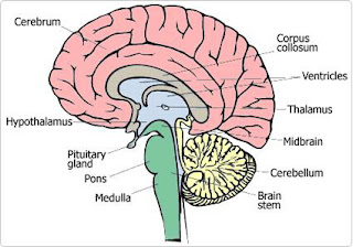
Your brain has three main components: the cerebrum (which consists of the left and right cerebral hemispheres), the cerebellum and the brain stem. The cerebral hemispheres of the brain make up the largest part of your brain. The cerebellum is the structure located behind the brain stem, and the brain stem is the lowest section of the brain and is connected to the spinal cord.
The central structures of the brain are the thalamus, hypothalamus, and pituitary gland. The thalamus relays sensory information to the cerebrum; the hypothalamus helps regulate body functions such as thirst and appetite, as well as sleep, aggression, and sexual behavior; and the pituitary gland produces hormones that play a role in growth, development, and various other physiological variables. The pons, medulla, and midbrain are the three structures that compose the brain stem. The ventricles are natural cavities inside the brain filled with cerebrospinal fluid.Respiratory System -- Basic Function
Your respiratory system provides the energy needed by cells of the body. Air is breathed in through the nasal cavity and/or mouth and down through the throat (the pharynx). The throat has three parts - the nasopharynx, the oropharynx, and the laryngopharynx. The air passes down the trachea (the windpipe), through the left and right bronchi, and into the lungs. Oxygen in the blood is delivered to body cells, where the oxygen and glucose in the cells undergo a series of reactions to provide energy to cells, and the waste product of this process is carried out of the lungs.
The larynx is your voice box; the epiglottis, a flap of cartilage that prevents food from entering the trachea; and the esophagus, the tube through which food passes to the stomach.Respiratory System -- Structure Detail
The Respiratory System - GlossaryBronchi: The two main air passages into the lungs.
Diaphragm: The main muscle used for breathing; separates the chest cavity from the abdominal cavity.
Epiglottis: A flap of cartilage that prevents food from entering the trachea (or windpipe).
Esophagus: The tube through which food passes from the mouth down into the stomach.
Heart: The muscular organ that pumps blood throughout the body.
Intercostal muscles: Thin sheets of muscle between each rib that expand (when air is inhaled) and contract (when air is exhaled).
Larynx: Voice box.
Lungs: The two organs that extract oxygen from inhaled air and expel carbon dioxide in exhaled air.
Muscles attached to the diaphragm: These muscles help move the diaphragm up and down for breathing.
Nasal cavity: Interior area of the nose; lined with a sticky mucous membrane and contains tiny, surface hairs called cilia.
Nose hairs: Located at the entrance of the nose, these hairs trap large particles that are inhaled.
Paranasal sinuses: Air spaces within the skull.
Pharynx: The throat.
Pleural membrane: Covering the lung and lining the chest cavity, this membrane has 2 thin layers.
Pulmonary vessels: Pulmonary arteries carry deoxygenated blood from the heart and lungs; pulmonary veins carry oxygenated blood back to the heart.
Respiratory center: Area of the brain that controls breathing.
Ribs: Bones attached to the spine and central portion of the breastbone, which support the chest wall and protect the heart, lungs, and other organs in the chest.
Trachea: Tube through which air passes from the nose to the lungs (also known as the windpipe).
Breast -- Disorders
The breasts can develop various types of disorders. Not all changes or lumps in the breast tissue mean that you have breast cancer. In fact, the vast majority of breast conditions are not cancerous. But if you find an abnormality or notice any changes, you should talk to your doctor. Some of the commonly occurring types of breast disorders are illustrated in the drawing below.
Muscles -- Back View
Each of your muscles is made up of thousands of thin, long, cylindrical cells called muscle fibers. The muscle fibers' highly specialized structure enables the muscles to relax and contract to produce movement. Muscles vary greatly in their shape and size, depending on their function.
Muscles -- Front View
Each of your muscles is made up of thousands of thin, long, cylindrical cells called muscle fibers. The muscle fibers' highly specialized structure enables the muscles to relax and contract to produce movement. Muscles vary greatly in their shape and size, depending on their function.
Circulation -- General
Your circulatory system consists of your heart and blood vessels. Together, these provide a continuous flow of blood to your body, supplying the tissues with oxygen and nutrients. Arteries carry blood away from the heart; veins return blood to the heart.
Circulation -- Arterial
Your arteries carry blood away from the heart. Oxygenated blood is pumped out of the heart through the body's main artery — the aorta. Arteries that branch off the aorta transport blood throughout the body, supplying tissues with oxygen and nutrients.
Circulation -- Venous
Your veins carry blood back toward the heart. Tiny vessels called capillaries in organs and tissues of the body deliver deoxygenated blood into small veins called venules, which join to form veins. Blood flows through the veins to the body's two main veins (called the vena cavae), which deliver the blood back into the heart.
Nervous System -- Basic
The brain and spinal cord comprise your central nervous system. The network of nerves that connect at different levels of the spinal cord control both conscious and unconscious activities. It is through the spinal cord that information flows from these nerves to the brain and back again.
Nervous System -- Groups of Nerves
Your nervous system is composed of the central nervous system, the cranial nerves, and the peripheral nerves. The brain and spinal cord together form the central nervous system. The cranial nerves connect the brain to the head. The four groups of nerves that branch from the cervical, thoracic, lumbar, and sacral regions of the spinal cord are called the peripheral nerves.
Digestive System
Your digestive system consists of organs that break down food into components that your body uses for energy and for building and repairing cells and tissues.
Food passes down the throat, down through a muscular tube called the esophagus, and into the stomach, where food continues to be broken down. The partially digested food passes into a short tube called the duodenum (first part of the small intestine). The jejunum and ileum are also part of the small intestine. The liver, the gallbladder, and the pancreas produce enzymes and substances that help with digestion in the small intestine.The last section of the digestive tract is the large intestine, which includes the cecum, colon, and rectum. The appendix is a branch off the large intestine; it has no known function. Indigestible remains of food are expelled through the anus.
Ear
The small cavity between the eardrum and inner ear conducts sound to the inner ear by three tiny bones called the malleus (the hammer), the incus (the anvil), and the stapes (the stirrup). The inner ear contains the cochlea (a coiled structure responsible for hearing), the semicircular canals (concerned with balance), and the vestible. The vestibule is an oval cavity that contains the saccule and utricle, which communicate with the cochlea and semicircular canals. The vestibular nerve passes impulses from the inner ear to the brain and is associated with balance; the cochlear nerve - part of the vestibular nerve - is associated with hearing.
Endocrine System
Your endocrine system is a collection of glands that produce hormones that regulate your body's growth, metabolism, and sexual development and function. The hormones are released into the bloodstream and transported to tissues and organs throughout your body. The Table below the illustration describes the function of these glands.
Skull
Your skull is composed of many bones that enclose the brain and form your facial skeleton.
Front | Side |
Skin
The skin — the largest organ of the body — is made up of a thin outer layer (called the epidermis) and a thicker outer layer (called the dermis). Below the dermis is the subcutaneous tissue, which contains fat. Buried in the skin are nerves that sense cold, heat, pain, pressure, and touch. Sebaceous glands secrete a lubricating substance called sebum. Deep within the skin are your sweat glands, which produce perspiration when you are too hot
Female Reproductive Organs
The female reproductive organs include the vagina (a muscular passage that connects the cervix with the external genital organs - one of which is a sensitive mound of tissue called the clitoris), the cervix (the lower part of the uterus that separates the body of the uterus from the vagina), the uterus (a hollow, muscular structure), the ovaries (two glands that produce certain hormones and contain tissue sacs in which eggs develop), and fallopian tubes (two muscular channels that connect the ovaries with the uterus). Fingerlike projections called fimbriae (located at the opening of the fallopian tubes) sweep an egg released from an ovary into the tube.
Female Reproduction -- Pregnancy
During pregnancy, a woman's body goes through a series of physical changes. At 6 months: the uterus has enlarged - and now extends above the level of the navel - to accommodate the growing fetus; the enlargement of the uterus displaces the abdominal organs upward. At 9 months: the abdominal organs continue to be pushed upward by the expanding uterus, and the fetus drops lower into the pelvis.
Month6 | Month9 |
Urinary System
Your urinary tract is the body system involved in the formation and excretion of urine. The kidneys filter out waste products from the blood. These waste products in combination with water are urine. The urine passes out of the kidneys through two narrow, muscular tubes called ureters. The ureters empty the urine into the bladder, and the urine is then excreted from the body through a tubelike structure called the urethra.


No comments:
Post a Comment